ARTÍCULO ORIGINAL
Effects of spermicidal extracts of Ananas comosus (pineapple) and Sapindus saponaria (jaboncillo) on cell viability, cytotoxicity and apoptosis
Efecto del extracto espermicida de Ananas comosus y Sapindus saponaria sobre la viabilidad, la citotoxicidad y la apoptosis celular
Mblga. Luisa Ospina Medina,I MSc. Ángela Álvarez Gómez,I MSc. Fabian Cortés Mancera,II MD. Ángela Cadavid Jaramillo,I Prof. Walter Cardona MayaI
I University of Antioquia, Medellín,
Colombia.
II Metropolitan Institute of
Technology. Medellín, Colombia.
Introduction: there are natural
products from different fruits and plants that are effective as spermicides,
but it is important that they should have little or no cytotoxic effect on epithelial
cells. Currently available spermicides with nonoxynol-9 cause vaginal irritation
and damage to the vaginal mucosa, the uterine epithelium, and the microbial
flora of the vagina.
Objective: to elucidate the effect on cell viability, cytotoxicity and apoptosis
of spermicidal extracts of Ananas comosus (L.) Merr. and Sapindus
saponaria L. over HeLa cell line.
Methods: both extracts were evaluated on HeLa cell line using the novel
ApoTox-Glo™ Triplex Assay to determine whether cell viability, cytotoxicity
and apoptosis were affected.
Results: it was determined that treatment with Sapindus saponaria
and Ananas comosus extracts initially affected cell viability, but the
latter tended to be restored. There was a sign of cell apoptosis that also tended
to decrease over time.
Conclusions: extracts of Sapindus saponaria and Ananas comosus
may affect the survival of cells at the beginning, but these can continue replicating
over time. There was a sign of cell apoptosis that also tended to decrease over
time. Something similar happened to cell cytotoxicity, indicating that although
the extracts may affect the survival of cells at the beginning (6 hours of treatment),
these can continue dividing over time.
Key words: natural compounds, spermicides, cell cytotoxicity, cell viability, cell apoptosis, Ananas comosus, Sapindus saponaria.
Introducción: diversos compuestos
de procedencia natural como frutos y plantas son altamente efectivos como espermicidas,
pero es necesario que estos no tengan efecto citotóxico sobre las células
epiteliales. Los espermicidas disponibles actualmente sobre la base de nonoxinol-9,
causan irritación y daño en la mucosa, el epitelio uterino y la
flora microbiana de la vagina.
Objetivo: determinar el efecto sobre la viabilidad, citotoxicidad y apoptosis
celular de extractos con actividad espermicida de Ananas comosus (L.)
Merr. y Sapindus saponaria L. sobre la línea celular HeLa.
Métodos: ambos extractos se evaluaron sobre la línea celular
HeLa para determinar el efecto en la viabilidad, la citotoxicidad y la apoptosis
celular, utilizando el novedoso ensayo triple ApoTox-Glo™.
Resultados: inicialmente el tratamiento con los extractos de Sapindus
saponaria y Ananas comosus afectaron la viabilidad celular; sin embargo,
esta tendió a restablecerse y mantenerse en el tiempo. Asimismo, la señal
de apoptosis celular tendió a disminuir a través de los tiempos
de tratamiento.
Conclusiones: los extractos de Sapindus saponaria y Ananas comosus
podrían afectar la viabilidad celular inicialmente; sin embargo, estas
continúan incrementándose con el paso del tiempo. Al mismo tiempo
la señal de apoptosis celular disminuyó a través del tiempo
y algo similar sucedió con la citotoxicidad celular, indicando que con
el paso de las horas los extractos pueden afectar la proliferación celular
al inicio (6 h de tratamiento), pero continúan proliferando.
Palabras clave: compuestos naturales, espermicidas, citotoxicidad celular, viabilidad celular, apoptosis celular, Ananas comosus, Sapindus saponaria.
INTRODUCTION
Among the many existing contraception methods, spermicides are one of the most interesting options due to advantages such as low cost, management by women and easy accessibility.1,2 There are natural products from different fruits and plants that are effective as spermicides that can kill or immobilize sperm cells.3-13 New spermicides should have little or no cytotoxic effect on epithelial cells and may act as microbiocides against pathogens. These aspects are a priority for future investigations because spermicides with nonoxynol-9 cause vaginal irritation and damage to the vaginal mucosa, the uterine epithelium,14-18 and the microbial flora of the vagina.19
After a review of the literature about the possible use of spermicides from Colombian plants,20 some studies have been carried out by our group where the effect of some of them has been reported.3-5,13,21 Using Passiflora edulis Sims (passion fruit), an immediate spermicidal effect and no cytotoxic effect were found on VERO and MDBK cells.3 In the case of Sapindus saponaria L. extract an immobilizing effect was found at five minutes without any cytotoxic effect on HeLa cells.13 Finally, sperm motility was affected after five minutes of incubation with Ananas comosus (pineapple) (L.) Merr. extract.5
The aim of this study was to test cell viability, cytotoxicity and apoptosis on HeLa cells after treatment with Ananas comosus and Sapindus saponaria extracts using ApoTox-Glo™ Triplex Assay.
METHODS
HeLa cells were plated in 96-well micro plates (3 x 10³ cells/well)22 in RPMI-1640 medium (GIBCO®, USA) with L-glutamine, supplemented with Fetal Bovine Serum 10 %, (FBS, GIBCO®, USA), penicillin streptomycin 1 % (GIBCO®, USA) and gentamicin 1 % (GENFAR®, Colombia), and incubated for 12 h, 37 °C, 5 % CO2.
Cells were stimulated with the polar extract of S. saponaria at 50 %,13 and the A. comosus extract at 50 % as the most spermicidal fraction and 12.5 %, which promoted a hyperactivation process on sperm cells5. Hydrogen peroxide 30 % (J. T. Baker Inc., Phillipsburg. N.J. USA) was used as a positive control of cytotoxicity and promoter of cellular apoptosis. RPMI without FBS was used as a negative control.
The cells were incubated for 6, 12, and 24 h at 37 °C, 5 % CO2, and then 10 µL of viability/cytotoxicity reagent containing both glycylphenylalanyl-aminofluorocoumarin (GF-AFC) substrate and bis-alanylalanyl-phenylalanyl-rhodamine 110 (bis-AAF-R110) substrate were added to all wells. After one hour of incubation at 37 °C the Relative Fluorescence Units for each signal were determined (505 nm excitation/400 nm emission for cell viability and 520 nm excitation/485 nm emission for cytotoxicity). Then, 10 µL of caspase reagent were added, and the Relative Luminiscence Units from the caspase activation were measured after 30 min of incubation. In all cases the GloMax-Multi Detection System (Promega) equipment was used. Each test of viability, cytotoxicity and apoptosis was run in triplicate.
The ApoTox-Glo™ Triplex Assay technique is based on the combination of three Promega assay chemistries to assess cell viability, cytotoxicity and caspase activation in a single assay well. The viable and metabolically active protease synthesizes a protease molecule of living cells specific to this cell type. If the molecule reaches the extracellular environment by loss of membrane integrity it reverts to its inactive state. This can be determined by applying the peptide substrate GF-AFC. When it enters the cell it is then cleaved by an enzyme reaction generating a fluorescence signal proportional to the number of viable cells. On the other hand, cells in a cytotoxic environment synthesize the dead cell protease that is found in the extracellular environment when the cells lose membrane integrity. This can also be determined or measured by applying the peptide substrate bis-AAF-R110. When the enzyme reaction occurs it generates a product that can also be measured by fluorescence. Both substrates and products are different and can be measured simultaneously. In the case of cells that are in a pro-apoptotic environment, activation of caspase 3/7 is determined by the addition of a substrate containing a tetrapeptide sequence DEVD. When the enzymatic reaction occurs, a luminescence signal forms that is directly proportional to the amount of caspase 3/7 produced in the cell (Promega Corporation. ApoTox-Glo™ Triplex Assay. Instructions for the use of products G6320 and G6321. Madison, USA).23
Statistical analysis
Mean values for each measurement of viability, cytotoxicity and apoptosis were calculated. Results are expressed in Relative Fluorescence Units (RFU) for viability and cytotoxicity, and in Relative Luminescence Units (RLU) for cell apoptosis. The data were analyzed using Microsoft Excel and Prism® software.
RESULTS
Fluorescence and luminescence signals in the plates that contain treated cells with hydrogen peroxide -positive control for cytotoxicity and apoptosis- were higher than the values expressed by cells treated with other stimuli, e.g. RPMI. In the case of viability, the fluorescence signal was weaker than the minimum values expressed by cells treated with other stimuli, e.g. RPMI and extracts.
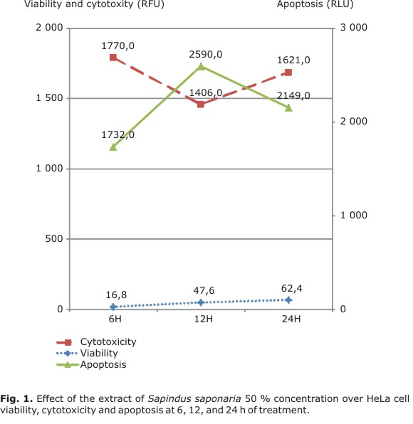
Under the treatment with the extract of S. saponaria, the signal of cell viability increased over the hours while the signal of apoptosis only increased until 12 h of treatment and then decreased at 24 h. On the other hand, the signal of fluorescence for cytotoxicity decreased after 12 h of treatment but it increased at 24 h (Fig. 1).
Treatment of cells with the supernatant of the extract of A. comosus affected cell viability at 6 and 12 h post-treatment but then at 24 h it tended to stabilize. The fluorescence signal, indicative of cytotoxicity, decreased at 12 h of treatment and then remained constant until 24 h. Cell viability decreased in this case due to the apoptotic effect of the plant extract. This was evidenced by the increase in Luminescence Relative Units at 6 and 12 h of treatment (Fig. 2).
In the case of the treatment with the extract of A. comosus 12.5 %, the signal of cell cytotoxicity was very similar over time. For cell apoptosis the strongest signal was at 12 h post-treatment and declined after 24 h. Similar results were found for cell viability (Fig. 3).
DISCUSSION
The test using in vitro assay cell models has become very important and its use is currently on the increase. Even cultures from vaginal or cervical tissues have been used as tools to determine the toxicity of some products, including spermicides.23 Therefore, our group chose to use a HeLa cell model in this study to determine the effect of the different plant extracts.
The results of this study are in line with those previously described regarding the treatment of HeLa cells with the extract of S. saponaria and A. comosus using the MTS assay, in which a compound of tretrazolium is added and biologically reduced to formazan by metabolically active cells, a soluble compound in the cell medium that is equal or proportional to the number of viable cells (Promega Corporation. CellTiter 96® AQueous Non-Radioactive Cell Proliferation Assay). In that case, it was only determined whether the cells entered into a cytotoxic environment due to any stimulus. Although that assay contributed very important results to this study, we determined whether cell viability was affected by a pro-apoptotic or cytotoxic process using the ApoTox-Glo™ Triplex Assay, which allowed a more complete understanding of what could happen in vivo. This way we could complement the results previously reported. As mentioned above, in these trials it was determined that treatment with S. saponaria and A. comosus extracts initially affected cell viability, but the latter tended to be restored. At the same time, there was a sign of cell apoptosis that also tended to decrease over time. Something similar happened to cell cytotoxicity, indicating that although the extracts of plants may affect the survival of cells at the beginning (6 h of treatment), these can continue replicating over time.
The use of techniques like the one presented in this study, which combines various reagents and measures three aspects of cellular activity, can mimic the complex cellular systems24 and can also give results based on what would happen in vivo.
ACKNOWLEDGEMENTS
This study was supported by the University of Antioquia (Sustainability 2011-2012) and the Metropolitan Institute of Technology (ITM), Medellín, Colombia. LOM, AAG and WCM were supported by a fellowship from Colciencias, Colombia.
REFERENCES
1. French RS, Cowan FM. Contraception for adolescents. Best Pract Res Clin Obstet Gynaecol. 2009;23(2):233-47.
2. Uribe-Clavijo M, Ospina-Medina L, Álvarez Gómez AM, Cortes-Mancera FM, Cadavid Jaramillo A, Cardona Maya W. Espermicidas: una alternativa de anticoncepcion para considerar. Tecnológicas. 2012;28:129-45.
3. Álvarez Gómez AM, Cardona Maya W, Forero J, Cadavid A. Human Spermicidal Activity of Passiflora edulis Extract. J Reproduction Contraception. 2010;21(2):95-100
4. Gallego G, Dubier H, Ospina L, Alvarez Gomez A, Arango V, Cardona Maya W, et al. Efecto de cinco extractos de plantas colombianas sobre espermatozoides humanos. Rev Cubana Plant Med. 2012;17(1):84-92.
5. Uribe-Clavijo M, Alvarez Gomez A, Arango V, Cortes-Mancera FM, Cadavid Jaramillo A, Cardona Maya W. Efecto in vitro del extracto vegetal de Ananas comosus sobre espermatozoides humanos. Tecnológicas. 2012;28:55-70.
6. Tso W, Lee C. Cottonseed oil as a vaginal contraceptive. Arch Androl. 1982;8(1):11-4.
7. Rajasekaran M, Nair AG, Hellstrom WJ, Sikka SC. Spermicidal activity of an antifungal saponin obtained from the tropical herb Mollugo pentaphylla. Contraception. 1993;47(4):401-12.
8. Souad K, Ali S, Mounir A, Mounir TM. Spermicidal activity of extract from Cestrum parqui. Contraception. 2007;75(2):152-6.
9. Kumar S, Chatterjee R, Dolai S, Adak S, Kabir SN, Banerjee S, et al. Chenopodium album seed extract-induced sperm cell death: exploration of a plausible pathway. Contraception. 2008;77(6):456-62.
10. Saha P, Majumdar S, Pal D, Pal BC, Kabir SN. Evaluation of spermicidal activity of MI-saponin A. Reprod Sci. 2010;17(5):454-64.
11. Anuja MM, Nithya RS, Swathy SS, Rajamanickam C, Indira M. Spermicidal action of a protein isolated from ethanolic root extracts of Achyranthes aspera: an in vitro study. Phytomedicine. international j phytotherapy phytopharmacology. 2011;18(8-9):776-82.
12. Paul S, Kang SC. In vitro determination of the contraceptive spermicidal activity of essential oil of Trachyspermum ammi (L.) Sprague ex Turrill fruits. New biotechnology. 2011;28(6):684-90.
13. Ospina L, Álvarez-Gomez A, Arango V, Cadavid A, Cardona-Maya W. Evaluación de la actividad espermicida y citotóxica del extracto de Sapindus saponaria L. (jaboncillo). Rev Cubana Plant Med. 2013;18(2).
14. Halpern V, Rountree W, Raymond EG, Law M. The effects of spermicides containing nonoxynol-9 on cervical cytology. Contraception. 2008;77(3):191-4.
15. Jain JK, Li A, Minoo P, Nucatola DL, Felix JC. The effect of nonoxynol-9 on human endometrium. Contraception. 2005;71(2):137-42.
16. Jain JK, Li A, Nucatola DL, Minoo P, Felix JC. Nonoxynol-9 induces apoptosis of endometrial explants by both caspase-dependent and -independent apoptotic pathways. Biol Reprod. 2005;73(2):382-8.
17. Stafford M, Ward H, Flanagan A. Safety study of N-9 as a vaginal microbicide: evidence of adverse effects. J Acquir Immune Defic Syndr Hum Retrovirol. 1998;17:327-31.
18. Watts DH, Rabe L, Krohn MA, Aura J, Hillier SL. The effects of three nonoxynol-9 preparations on vaginal flora and epithelium. J Infect Dis. 1999;180(2):426-37.
19. Ojha P, Maikhuri JP, Gupta G. Effect of spermicides on Lactobacillus acidophilus in vitro-nonoxynol-9 vs. Sapindus saponins. Contraception. 2003;68(2):135-8.
20. Álvarez Gómez A, Cardona Maya W, Castro Álvarez J, Jimenez S, Cadavid A. Nuevas opciones en anticoncepción: posible uso espermicida de plantas colombianas. Actas Urol Esp. 2007;31(4):372-81.
21. Puerta Suárez J, Cardona Duque D, Álvarez Gómez A, Arango V, Cadavid A, Cardona Maya W. Efecto del extracto de Anethum graveolens, Melissa officinalis y Calendula officinalis sobre espermatozoides humanos. Rev Cubana Plant Med. 2012;17(4):420-30.
22. Sadeghi Aliabadi H, Ghasemi N, Kohi M. Cytotoxic effect of Convolvulus arvensis extracts on human cancerous cell line. Research Pharmaceutical Sciences. 2008;3(1):31-4.
23. Ayehunie S, Cannon C, Larosa K, Pudney J, Anderson DJ, Klausner M. Development of an in vitro alternative assay method for vaginal irritation. Toxicology. 2011;279(1-3):130-8.
24. Niles A, Moravec R, Eric Hesselberth P, Scurria M, Daily W, Riss T. A homogeneous assay to measure live and dead cells in the same sample by detecting different protease markers. Anal Biochem. 2007;366(2):197-206.
Recibido: 10 de septiembre de 2012.
Aprobado: 23 de marzo de 2013.
Walter Cardona Maya. Grupo Reproducción, Facultad de Medicina, Universidad de Antioquia, Calle 52 # 61-30, Laboratorio 534. Phone: (574) 2196476. E-mail: wdcmaya@medicina.udea.edu.co; wdcmaya@gmail.com